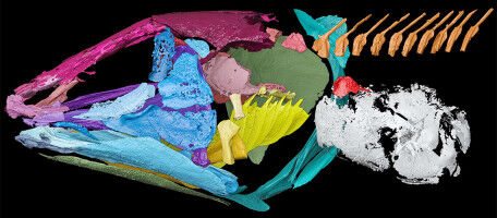
© 2020 EPFL - EPFL biophysicists have developed a high-throughput super-resolution microscope to probe nanoscale structures and dynamics of mammalian cells, showing in unprecedented detail the twists and turns of an organelle important for cell division. If you want to understand the underlying mechanisms of cellular motility and division, then the centriole is the organelle of interest. Each cell has a pair of centrioles which help to segregate chromosomes during cell division. These special organelles are multi-molecular machines composed of hundreds of proteins and have a hidden code of post-translational modifications (PTMs), that contribute to their rigidity or flexibility, which in turn may help explain how centrioles function. Based on previous studies mostly using electron microscopy, the basic structure of centrioles is known. But PTMs are invisible to the electron microscope, so what do they look like? Thanks to improved super resolution fluorescence microscope technology developed by EPFL biophysicists, we now have a detailed picture of these nanoscale structures, both isolated and in situ . As expected, the centrioles are shaped like ridged bullets, i.e. they are cylindrical with nine lengthwise ridges and their diameter tapers off at one end.
TO READ THIS ARTICLE, CREATE YOUR ACCOUNT
And extend your reading, free of charge and with no commitment.
Your Benefits
- Access to all content
- Receive newsmails for news and jobs
- Post ads